WWW.Cell Biology Education
Each quarter, Cell Biology Education calls attention to several Web sites of educational interest to the life science community. The journal does not endorse or guarantee the accuracy of the information at any of the listed sites. If you want to comment on the selections or suggest future inclusions, please send a message to [email protected]. The sites listed below were last accessed on June 1, 2003.
CANCERQUEST
http://cancerquest.org/
Many biologists have only an indirect knowledge of the cancer biology field. The CancerQuest site gives a very clean overview of the biology of cancer and at the same time provides a wonderful structure for teaching the topic to either high-school or college audiences. Dr. Gregg Orloff of the Emory University Biology Department developed the concept and design of the site. Working with students and Emory IT (Information Technology) professionals, the effort has received funding through a Hughes Science Initiative and with Emory resources.
Figure 1. The introductory screen logo for CancerQuest. Graphic used with the permission of Gregg Orloff, Emory University.
The site is divided into 12 topics, with a topic having as many as 10 subsections. The subsections are divided into individual parts, each of which is displayed on two or fewer less computer pages. (See Figure 1, where subsections for each topic are listed under the title screen.) Some parts have computer animations to clarify the concept. (See Figure 2 for a ribozyme cleavage image.) The animations require the free software Flash 6 player in order to operate. (Flash 6 player found at http://www.macromedia.com/.) Each subsection part is tightly focused and easy to grasp by the viewer. Within the text are hypertext words; when selected, the word appears in dictionary style with definitions. Also within the text are arrow icons that lead to very current references for key statements, a very nice feature.
The first four topics deal with common biology material: biological molecules (with molecular rotations), cell structure, central dogma, and cell division. Although these topics are meant to support the cancer biology information to follow, the presented material would complement many biology courses. Transitioning into the specific cancer material are two topics on genetics. Here the concepts of oncogenes and tumor suppressors are beautifully animated. DNA mutation and underlying causes are nicely explored as well.
Figure 2. An example of a ribozyme animation graphic. The concept being demonstrated has to do with methods of cancer treatment. Graphic used with the permission of Gregg Orloff, Emory University.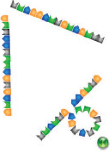
The next three topics get at the heart of the CancerQuest site: tumor biology, diagnosis, and treatment. Terms like carcinoma are carefully defined. Although the word carcinoma is familiar to many, few realize that a carcinoma cell is derived from epithelial cells and constitute about 85% of the reported cancers. Figure 3 demonstrates how enzymes from carcinoma cells allow the cancer cells to escape from the epithelial surface and enter the depths of the body. Tumor biology and metastasis are explored in an understandable way, with references given to other sources of information such as a Public Broadcasting System's Nova special on cancer.
The topic title “Diagnosis and Detection” is exceptionally well done. Colonoscopy is explained, with very clear illustrations of what normal, polyp, and cancerous tissue are. How tumors are staged is clearly outlined as is an extremely useful section demonstrating how to read a pathology report. Again, the topic has words with which most people are familiar but that have never put together in such a clear fashion. Links are provided to quality sources of information.
Figure 3. An illustration from an animation demonstrating how matrix metalloproteases carve through the basal lamina and allow carcinoma cells to escape into the bloodstream. Graphic used with the permission of Gregg Orloff, Emory University.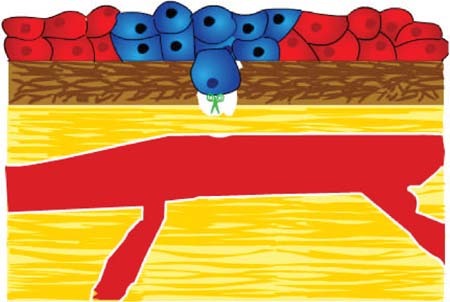
The next topic explores treatment and contains over 75 pages of information. Often as biologists we are asked how chemotherapy works. Rarely do we have the background to answer specific questions. This treatment section provides a broad understanding of current treatment modalities. The presentation sequence is superb. References and links to more information are provided. Readers can gather a broad perspective about cancer treatment in a very compact and understandable format.
The last two topics direct the Web viewer to finding information about clinical trials and additional references. There is a search engine built into the site. CancerQuest is an impressive site with a high value for teaching as well as for personal information gathering. Be prepared to spend some time at this URL. Hats off to Dr. Orloff, his staff, and Emory University for developing a very needed and thoughtful site.
Figure 4. The exercise is based on a Bausch & Lomb Galen II compound microscope. Graphic used with the permission of Robert Ketcham, University of Delaware.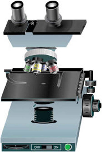
VIRTUAL MICROSCOPE
http://www.udel.edu/Biology/ketcham/microscope/
In a world full of electronic gadgets and widgets of all kinds, many students fail to grasp the fundamentals of operating a standard optical light microscope. Robert Ketcham and Becky Kinney of the University of Delaware have developed an interactive Web program that allows students to practice using a microscope over the Internet. Working with university funding, the Virtual Microscope lab has been produced primarily for nonmajors taking their initial biology laboratory.
Ketcham and Kinney base their microscopy lab exercise on the Bausch & Lomb Galen II compound microscope, a widely used student microscope (Figure 4). A student is instructed to practice systematically 10 elements of microscopy usage (Figure 5). Students are instructed to turn on the light, set the light rheostat, place the low-power lens in position, center the specimen, and move the stage to the top position. As obvious as this all seems to a practiced microscopist, many students have not picked up these fundamentals. The next sequence is to adjust the oculars, course focus, iris diaphragm, and fine focus. By using the computer program, novice microscope users can practice these maneuvers before they begin working with real microscopes in the lab.
Figure 5. A concept checklist for the use of the virtual microscope. Used with permission of Robert Ketcham, University of Delaware.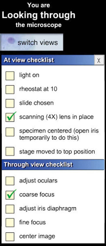
The current version of this digital-based microscope comes with four slides: the letter “e,” an onion root tip (Figure 6), a bacterial capsule, and a cheek smear. One can envision additional slides being added over time. Associated with the learning experience is a problem set that causes the students to examine”puzzles” such as an out-of-focus microscope. This virtual microscope comes with an ocular micrometer and the problem set also includes some measurement exercises. Complementing the virtual microscope is a 7-min video on how to use the microscope. The video can be played through a number of publicly available Web formats and in different screen sizes to accommodate various connection speeds.
Although this site is still a work in progress, it has great value in preparing a student for the first microscopy lab. It is both a fun site to visit and a perfect place to start practicing good microscopy habits.
Figure 6. The onion root tip slide as viewed through the virtual microscope. Used with permission of R. Ketcham, University of Delaware.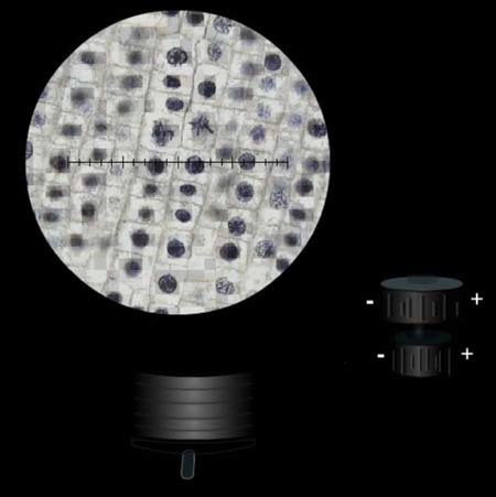
FOOTNOTES
Monitoring Editor: A. Malcolm Campbell


